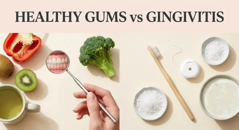
Healthy bottom gums are firm, pale to coral pink in color, and fit snugly against the base of each lower tooth without swelling, bleeding, or pulling away. Most people never think about their gum tissue until something feels off, and the lower arch is often where the first signs appear.
According to the American Academy of Periodontology (AAP), nearly half of American adults over 30 show some form of gum disease, and the mandibular (lower jaw) front teeth are among the most commonly affected areas due to plaque accumulation patterns and brushing habits. Getting a clear picture of what healthy actually looks like is the first step to protecting it.
This article covers the visual hallmarks of healthy lower gum tissue, the key differences between healthy and unhealthy gums, what bottom gum recession looks like, which nutrients directly support gum health, and what daily habits have the strongest evidence behind them.
Healthy Bottom Gums: What They Are and Why They Matter
Healthy bottom gums are the soft tissue, known clinically as the gingiva, that surrounds and supports the roots of the lower teeth along the mandibular arch.
The gingiva has two distinct zones. The marginal gingiva is the thin edge that meets the tooth at the gum line. The attached gingiva sits below that, firmer and bound to the underlying alveolar bone. Both zones need to be in good condition for the lower teeth to remain stable.
The lower front teeth, the mandibular incisors, are particularly vulnerable to gum changes. Their roots are closer to the outer bone surface, which means gum recession here can progress faster than elsewhere in the mouth. That’s why knowing what healthy looks like at this specific location matters more than a generic gum health overview.
The National Institute of Dental and Craniofacial Research (NIDCR) describes healthy gingival tissue as the foundation of long-term tooth retention. Without intact gum support, teeth lose the structural anchoring they depend on.
Key characteristics of healthy gum tissue include:
- Firm, not puffy or spongy, to the touch
- Pale to coral pink coloring (with natural variation by skin tone)
- Tight seal around each tooth base
- No bleeding during normal brushing or flossing
- No persistent soreness or sensitivity at the gum line
What Do Healthy Bottom Gums Look Like?
Healthy bottom gums look pale to coral pink, lie flat against each tooth, and show a slightly textured surface that dentists describe as stippled, resembling the surface of an orange peel.
That stippled texture is actually a sign of healthy collagen fiber organization within the tissue. Smooth, shiny, or puffy gums around the lower teeth can indicate early inflammation, even before pain appears. Healthy tissue doesn’t puff up between teeth or hang loosely over the tooth surface.
The gum line on the lower arch typically sits slightly higher above the roots than it appears in the upper jaw, because gravity and muscle movement both place different mechanical demands on the mandibular gingiva. A consistent, even gum margin at each lower tooth is the target.
The American Dental Association (ADA) notes that the gum line should form a smooth scalloped curve around each tooth, dipping slightly between teeth to form what’s called the interdental papilla. When this papilla looks blunted, red, or missing, that’s a clinical signal.
| Feature | Healthy Appearance | Concern Signal |
|---|---|---|
| Color | Pale to coral pink | Red, dark red, purple, or white patches |
| Texture | Slightly stippled (orange peel) | Smooth, shiny, or puffy |
| Gum margin | Tight, even scallop around each tooth | Uneven, receded, or rolled outward |
| Interdental papilla | Pointed, fills the space between teeth | Blunted, missing, or swollen |
| Bleeding | None during brushing or flossing | Any bleeding, even occasional |
| Soreness | None at rest or during normal function | Sensitivity to touch or pressure |
Normal Healthy Bottom Teeth: What the Full Picture Includes
Normal healthy bottom teeth are those supported by stable, attached gum tissue, with no visible root exposure and no changes in how the teeth fit together when you bite.
The lower arch typically contains 14 to 16 teeth in an adult mouth, including incisors, canines, premolars, and molars. Each tooth’s root is partially encased in alveolar bone and anchored to it by the periodontal ligament, a network of connective tissue fibers. The gum tissue overlying this system should be firmly attached and show no signs of detachment from the tooth surface.
A healthy lower tooth shows no darkening at the gum line, which would indicate root exposure. The crown of the tooth, the visible white portion, should not appear longer than normal. An elongated-looking lower tooth is often the first visual sign that gum recession has occurred.
Think of healthy gum tissue the way you’d think of the caulk sealing a kitchen sink: when it’s intact, nothing gets in. When it starts to pull away at the edges, the underlying structure becomes vulnerable to moisture, pressure, and eventual damage.
According to a 2019 review in the Journal of Periodontology, loss of clinical attachment in the lower anterior region is consistently associated with long-term tooth mobility if not addressed at the early stage.
Quick Tip:
- Check your lower front teeth in good lighting with a dental mirror once a month
- Look for consistent gum height at each tooth, not just color changes
- Slight gum line asymmetry between adjacent teeth warrants a professional assessment
Key Takeaway: Healthy bottom gums are firm, pale to coral pink, stippled in texture, and form a tight scalloped margin at each lower tooth. Any puffiness, bleeding, or change in the gum line height is worth professional attention.
Healthy Lower Teeth: The Role of Bone and Tissue Together
Healthy lower teeth depend on two structures working together: the gum tissue you can see and the alveolar bone underneath that you can’t.
The bone level around lower teeth is not visible to the naked eye. A dental X-ray is required to assess it properly. But gum health above the surface often reflects what’s happening below: when the bone recedes due to chronic bacterial infection, the gum tissue follows.
The NIDCR estimates that by age 65, more than 70% of adults have experienced some degree of bone loss around at least one tooth. Early investment in gum tissue health significantly slows that progression.
Lower teeth also face greater mechanical stress than upper teeth in most people. Nighttime grinding (bruxism) disproportionately affects the mandibular arch and directly damages the gingival attachment. People who grind their teeth often see localized gum recession at the lower canines and premolars first.
| Lower Tooth Region | Common Gum Issue | Early Warning Sign |
|---|---|---|
| Front incisors | Recession due to aggressive brushing | Visible root surface, tooth sensitivity |
| Canines | Recession from bruxism or crowding | Gum pulling away on the outer surface |
| Premolars | Plaque accumulation, early pocket formation | Slight swelling between premolars |
| Molars | Deep pocket formation | Bleeding at the back during flossing |
Healthy Bottom Teeth Gums: The Gum-Tooth Junction Explained
The gum-tooth junction, clinically called the sulcus, is the shallow groove where healthy gum tissue meets the tooth surface. In a healthy mouth, this groove measures between 1 and 3 millimeters deep.
A dentist or hygienist measures this depth during a periodontal probing examination. Numbers between 1 and 3 mm indicate health. Numbers of 4 mm or above indicate a pathological pocket where bacteria accumulate below the reach of a regular toothbrush.
Most people have no idea this groove exists until a hygienist calls out numbers during a cleaning. Hearing “3” repeatedly is good. Hearing “5” or “6” at the back lower molars is an early clinical alert.
The junction also matters for how you brush. Angling a soft-bristle toothbrush at approximately 45 degrees toward the gum line, using short circular or vibratory strokes, is designed specifically to clean this sulcular area. Brushing straight across the gum line, the most common technique, misses this zone entirely.
According to ADA clinical recommendations, brushing technique matters as much as brushing frequency for maintaining a healthy sulcular environment at the lower arch.
Key Tip:
- Ask your dental hygienist to tell you your probing numbers at your next cleaning
- Numbers of 4 mm or more at any lower tooth warrant a follow-up conversation about your current brushing and flossing technique
Healthy Gum Color: What Shades Are Normal and What Aren’t
Healthy gum color ranges from pale pink to coral pink for lighter skin tones, and from medium brown to deep brownish-pink in people with more melanin-rich skin. Both are completely normal.
This is one of the most misunderstood aspects of gum health. Medical diagrams and dental education materials have historically used a single standard of pale pink gum tissue, which does not reflect the natural variation across skin tones. Melanin pigmentation in gum tissue is normal, genetically determined, and completely unrelated to disease.
What matters is consistency. A person with naturally dark, evenly pigmented gum tissue has healthy gums if the color is uniform and stable. Sudden changes in gum color, especially patchy red or white areas, are the clinical signal, not the baseline color itself.
Colors that warrant attention regardless of skin tone:
- Bright red or deep red gums: often inflammation from plaque or bacteria
- Purple or bluish-tinged gums: can indicate reduced circulation or severe chronic inflammation
- White patches (leukoplakia): require professional evaluation, as causes range from irritation to tissue changes that need assessment
- Pale white or grayish gum tissue: may indicate reduced blood flow or, in rare cases, systemic conditions
A study published in the Journal of Clinical Periodontology in 2020 confirmed that racial and ethnic variation in gingival pigmentation is a normal physiological finding with no clinical pathological significance.
Key Takeaway: Gum color varies normally by skin tone and ethnicity. Focus on consistency and any sudden changes rather than comparing your gum color to a single textbook standard.
Healthy v Unhealthy Gums: A Side-by-Side Breakdown
The clearest way to distinguish healthy versus unhealthy gums is to compare specific clinical signs directly, rather than relying on a vague sense of “something looks off.”
Healthy gum tissue does not bleed. This is the single most reliable home indicator. The American Academy of Periodontology (AAP) states that bleeding on brushing is not normal and is the primary early indicator of gingivitis, the earliest reversible stage of gum disease.
Unhealthy gums often feel different before they look different. Puffiness around the base of a lower tooth, increased sensitivity to cold water at the gum line, or a persistent bad taste in the mouth can precede visible color changes by weeks.
| Sign | Healthy Gums | Unhealthy Gums |
|---|---|---|
| Color | Pink to brownish-pink (consistent) | Red, purple, white patches, or sudden change |
| Texture | Slightly stippled or firm | Smooth, shiny, puffy, or boggy |
| Bleeding | None | Bleeds on brushing, flossing, or light pressure |
| Gum margin | Tight at tooth base | Swollen, rolled, or receding |
| Odor | Neutral with good oral hygiene | Persistent bad breath despite brushing |
| Sensitivity | None at gum line at rest | Tender to touch, sensitivity to temperature |
| Pocket depth | 1 to 3 mm | 4 mm or greater |
| Gum attachment | Stable, no movement | Gum separating from tooth surface |
The good news: gingivitis, the early stage, is fully reversible. The ADA confirms that consistent plaque removal through brushing and interdental cleaning is sufficient to reverse gingivitis in most adults within 2 to 4 weeks.
Signs of Unhealthy Gums: When to Take Action
The most common signs of unhealthy gums are bleeding, persistent redness, puffiness between teeth, and gum recession that makes lower teeth appear longer than before.
Bleeding is the most misunderstood sign. Many people assume some bleeding during brushing is normal or means they’re brushing “too hard.” Bleeding gum tissue is inflamed tissue. Inflammation signals a bacterial response, not a mechanical injury from the brush.
Persistent bad breath that doesn’t resolve after brushing is another common early indicator. The sulfur-producing bacteria associated with periodontal infection produce volatile sulfur compounds (VSCs) that cause a distinct, persistent odor. Brushing the tongue and using water flossing can reduce VSC load, but they don’t address the underlying bacterial environment in infected pockets.
Signs that indicate you should schedule a dental assessment sooner rather than later:
- Bleeding that persists for more than 2 weeks of improved brushing and flossing
- Visible root exposure on any lower front tooth
- A tooth that feels loose or shifts slightly when pressed
- Pain when chewing that wasn’t present before
- Swelling that appears suddenly in the lower jaw area
- White or red patches on the gum tissue that last more than 2 weeks
According to the CDC, gum disease is the leading cause of tooth loss in adults in the United States. Early identification directly changes long-term outcomes.
Key Takeaway: Bleeding during brushing is not normal and not a sign of brushing too hard. It’s the most reliable early home indicator that gum tissue needs attention.
Healthy Gum Texture and Firmness: What You Should Feel
Healthy gum tissue feels firm and resilient when pressed gently with a finger. It doesn’t dimple, stay compressed, or feel spongy.
The texture term dentists use is stippling, which refers to the slightly dimpled surface visible on firmly attached gum tissue. Under magnification, this texture reflects well-organized collagen fiber bundles within the tissue. When inflammation develops, those fibers loosen and the tissue becomes edematous (swollen with fluid), producing the smooth, puffy texture of inflamed gums.
Gum firmness also depends on how well the tissue is keratinized. The attached gingiva, the portion bound to the bone below, is keratinized and therefore tougher. The alveolar mucosa below it is not keratinized and appears darker, shinier, and more delicate. The junction between these two tissue types is visible in the lower arch as a slight color change, and it’s a normal anatomical feature.
People who have undergone gum grafting procedures may find their bottom gums feel different in texture in the treated area, which is expected and not a sign of ongoing disease.
According to research published in the Journal of Periodontology, stippled texture in the attached gingiva is associated with well-organized collagen architecture and lower rates of periodontal pocket formation in longitudinal studies.
| Texture Sign | What It Indicates |
|---|---|
| Slightly stippled, firm | Healthy, well-organized connective tissue |
| Smooth and shiny, normal color | Early edema; possible mild gingivitis |
| Smooth, shiny, and red or purple | Active inflammation; clinical evaluation needed |
| Cratered or pitted between teeth | Previous or active tissue destruction |
| Fibrous and overgrown | Possible drug-induced gingival overgrowth or chronic irritation |
Bottom Gum Recession Signs: What to Watch For
Bottom gum recession means the gum tissue at the lower arch has pulled away from its original position at the tooth crown, exposing a portion of the root surface below. The first visible sign is a tooth that appears longer than it used to.
The lower front incisors are the most commonly affected teeth. Their anatomy, combined with aggressive brushing habits, toothbrush abrasion from hard bristles, and thin underlying bone, makes them the most recession-prone teeth in the mouth.
Root sensitivity is often the first symptom people notice before visible recession is apparent. Cold water, cold air, or sweet foods triggering a sharp, brief sensitivity at the base of a lower front tooth is a signal worth acting on early.
Common causes of bottom gum recession:
- Overly aggressive brushing technique or use of a medium or hard bristle brush
- Toothbrush abrasion from brushing immediately after acidic foods or drinks
- Chronic untreated gingivitis progressing to periodontitis
- Bruxism (night grinding) placing lateral forces on lower canines and incisors
- Dental crowding that positions roots outside the alveolar bone envelope
- Piercings or oral jewelry that physically traumatize the gum tissue
The AAP classifies gum recession into four clinical classes based on depth and tissue loss. Classes I and II are often amenable to conservative treatment and gum grafting. Classes III and IV involve bone loss and require more involved periodontal management.
A dentist can confirm recession during examination. If you suspect it at home, compare photographs of your lower teeth taken several months apart. A measurable change in visible tooth length is an objective marker.
Key Takeaway: Bottom gum recession often begins as tooth sensitivity before it’s visible. Switching to a soft-bristle toothbrush and adjusting brushing angle are two evidence-supported changes that slow its progression.
Foods That Help Gum Health
The foods most directly linked to gum tissue health are those that supply ascorbic acid (vitamin C), calcium, omega-3 fatty acids, and polyphenol antioxidants, all of which have documented roles in collagen synthesis and inflammatory regulation within periodontal tissue.
Vitamin C is the most well-established dietary factor in gum health. Ascorbic acid is a required cofactor for collagen synthesis. Without adequate vitamin C, the collagen matrix holding gum tissue together weakens. The NIH Office of Dietary Supplements recommends 65 to 90 mg of vitamin C daily for adults, with an upper limit of 2,000 mg. Smokers require an additional 35 mg daily due to increased oxidative stress.
Green tea (Camellia sinensis) is one of the more researched functional foods for periodontal health. Its polyphenols, particularly epigallocatechin gallate (EGCG), have been shown in multiple clinical trials to reduce gingival inflammation markers when consumed regularly.
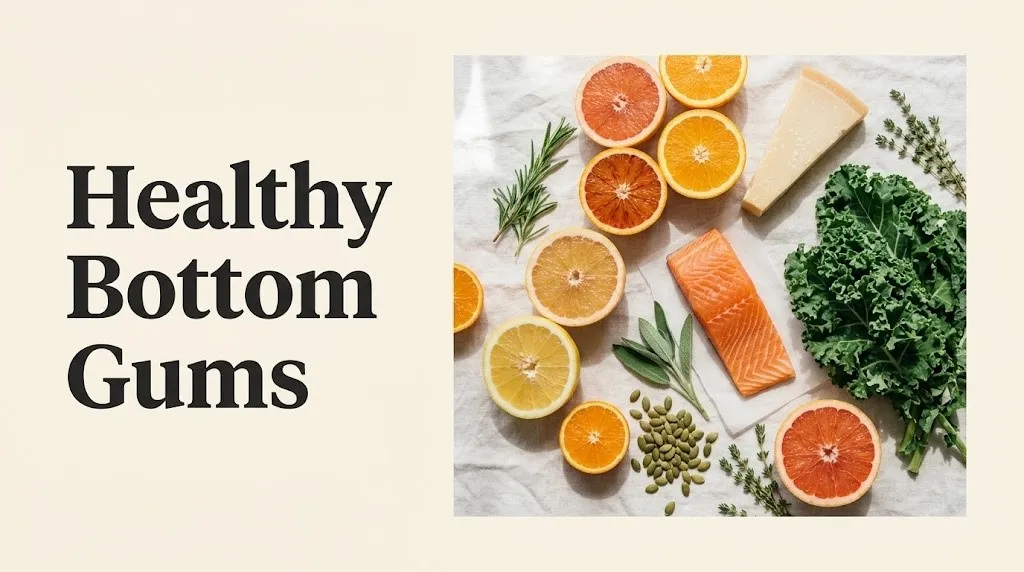
Foods that directly support gum tissue:
- Citrus fruits (oranges, kiwi, strawberries): ascorbic acid (vitamin C) for collagen production
- Leafy greens (spinach, kale, Swiss chard): calcium, vitamin K, and folate supporting tissue repair
- Fatty fish (salmon, mackerel, sardines): omega-3 fatty acids (EPA and DHA) for anti-inflammatory support
- Dairy products (plain yogurt, cheese, milk): calcium and casein phosphopeptides supporting tooth and gum mineral integrity
- Green tea: EGCG polyphenols associated with reduced gingival bleeding in clinical research
- Nuts and seeds (almonds, sunflower seeds): zinc and vitamin E supporting immune response in gingival tissue
- Carrots and celery: fibrous texture stimulates saliva production and mechanically reduces plaque
People with dairy allergies or lactose intolerance can substitute fortified plant-based milks (soy, oat, or almond, ensuring they contain added calcium) to maintain calcium intake without digestive discomfort.
Vitamins for Gum Health: What the Research Shows
The three vitamins with the strongest direct evidence for gum tissue health are vitamin C (ascorbic acid), vitamin D, and vitamin K2 (menaquinone).
Vitamin C is indispensable for gingival integrity. Its role in collagen cross-linking means that even subclinical deficiency, well above the level of scurvy, can impair gum tissue repair. A 2021 systematic review published in Nutrients found that patients with periodontal disease consistently showed lower serum vitamin C levels than periodontally healthy controls.
Vitamin D’s role in gum health is less discussed but well-supported. Vitamin D receptors are present in gingival fibroblasts and immune cells within the periodontium. The NIH recommends 600 IU of vitamin D daily for adults under 70. Research published in the Journal of Periodontology has linked vitamin D deficiency with increased rates of gingival inflammation and periodontal pocket formation.
| Vitamin | Daily Recommended Intake | Food Sources | Gum Health Role |
|---|---|---|---|
| Vitamin C (ascorbic acid) | 65 to 90 mg/day | Citrus, kiwi, strawberries, bell pepper | Collagen synthesis, tissue repair |
| Vitamin D | 600 IU/day | Fatty fish, fortified milk, egg yolk, sunlight | Anti-inflammatory, immune modulation |
| Vitamin K2 (menaquinone) | 90 to 120 mcg/day | Fermented foods (natto), cheese, egg yolk | Bone metabolism supporting periodontal bone |
| Zinc | 8 to 11 mg/day | Pumpkin seeds, beef, chickpeas | Wound healing, immune defense in gingival tissue |
| Omega-3 (EPA and DHA) | 250 to 500 mg/day combined | Salmon, sardines, mackerel, algae oil | Prostaglandin regulation, reduced gingival inflammation |
People taking anticoagulant medications like warfarin should confirm any increase in vitamin K-rich foods or supplements with their prescribing physician, as vitamin K directly affects clotting factor metabolism.
Key Takeaway: Vitamin C, vitamin D, and vitamin K2 have the strongest direct evidence for gum tissue health. Getting them through food first, then supplementing specific deficiencies with a healthcare provider’s guidance, is the most evidence-supported approach.
How to Keep Bottom Gums Healthy
Keeping bottom gums healthy comes down to four evidence-supported practices: consistent plaque removal with correct technique, interdental cleaning daily, avoiding habits that physically damage gum tissue, and maintaining adequate nutritional status for tissue repair.
Brushing technique is the most common area where people unknowingly cause harm. A medium or hard bristle brush, or an aggressive horizontal scrubbing motion, is a well-documented cause of gum recession at the lower front teeth. The ADA recommends a soft-bristle toothbrush angled at 45 degrees to the gum line, using a gentle circular motion rather than horizontal strokes.
Electric toothbrushes with pressure sensors offer a practical advantage here. They signal when you’re pressing too hard, which is the brushing behavior most directly associated with abrasion-related recession at the lower arch.
To establish a gum-protective brushing and cleaning routine:
- Choose a soft-bristle toothbrush or an oscillating-rotating electric brush with a pressure sensor
- Position the bristle head at a 45-degree angle to the gum line at the lower front teeth
- Use short, gentle circular or vibratory strokes for 30 seconds per quadrant
- Floss or use an interdental brush once daily, working gently beneath the contact point between each lower tooth
- After flossing, consider a 30-second rinse with a fluoride mouthwash if your dentist recommends it for your situation
- Wait at least 30 minutes after eating acidic foods (citrus, vinegar-based dishes, carbonated drinks) before brushing to allow enamel to remineralize before any mechanical stress
- Schedule a professional cleaning every 6 months, or every 3 to 4 months if your dentist has identified active periodontal pocketing at any lower tooth
Smoking is one of the most studied risk factors for gum disease progression. The CDC reports that smokers are twice as likely to develop gum disease as non-smokers, and that smoking masks the bleeding response, making the visual signs of disease harder to detect.
What Do Healthy Bottom Teeth Look Like: The Full Visual Guide
Healthy bottom teeth look white to slightly off-white in crown color, with no visible darkening at the gum line, and are supported by gum tissue that sits evenly at the base of each tooth without recession or swelling.
The crown of a lower tooth should not look noticeably longer than the tooth next to it. If one lower front tooth looks visibly longer, that’s a sign its gum margin has receded. Healthy crowns are roughly uniform in visible length along the lower arch.
The gum surface between lower teeth should fill the space between the crowns with a pointed triangular papilla. When you look at the spaces between your lower front teeth, a filled-in pointed tip of pink tissue is a healthy sign. A flat, missing, or rounded-off papilla between lower front teeth can indicate previous gum tissue loss, often from untreated gingivitis or aggressive flossing technique.
A healthy lower arch also shows no calculus (tartar) deposits visible as yellowish or brownish buildup at the gum line. Calculus is hardened mineralized plaque that only a dental scaler can remove. Its presence at the lower front teeth (one of the most common tartar accumulation sites, due to proximity to the salivary gland ducts beneath the tongue) indicates that brushing alone isn’t removing plaque effectively in this area.
| Feature | Healthy Lower Teeth | Concern Indicator |
|---|---|---|
| Crown color | White to slightly off-white | Dark margins at gum line (root exposure or decay) |
| Crown length | Uniform across adjacent teeth | One tooth noticeably longer (recession) |
| Interdental papilla | Pointed, fills the contact space | Flat, missing, or blunted |
| Gum line | Even scallop, no gaps | Uneven margins, visible root surface |
| Calculus | None visible | Yellowish-brown deposits at gum line |
| Crowding | Teeth align with accessible contact points | Overlapping crowns making interdental cleaning difficult |
Key Takeaway: Healthy bottom teeth show uniform crown length, filled-in interdental papillae, and no visible tartar buildup at the gum line. A change in how long your lower front teeth appear is the clearest home signal of gum recession.
Frequently Asked Questions About Healthy Bottom Gums
What color should healthy bottom gums be?
Healthy bottom gums range from pale pink to coral pink in lighter skin tones, and from medium brown to dark brownish-pink in people with higher melanin levels.
Both are completely normal and reflect natural genetic variation in melanin pigmentation.
The key indicator of health is color consistency: stable, even color with no sudden red, white, or purple patches.
Why are my bottom gums receding?
Bottom gum recession most often results from aggressive brushing technique, a medium or hard bristle toothbrush, or chronic untreated gum inflammation.
Bruxism (nighttime grinding), dental crowding, and oral jewelry that contacts the lower front gum tissue are additional documented causes.
A dentist can identify the specific contributing factor during examination and recommend targeted treatment before significant tissue loss occurs.
How do I know if my lower gums are unhealthy?
The most reliable home indicators of unhealthy lower gums are bleeding on brushing or flossing, persistent puffiness at the gum line, and lower front teeth that appear longer than they previously did.
Any new sensitivity to cold or pressure at the base of a lower tooth is worth professional evaluation.
Symptoms lasting more than two weeks despite improved brushing and flossing should prompt a dental assessment.
What foods help keep gums healthy?
Foods highest in ascorbic acid (vitamin C), calcium, and omega-3 fatty acids have the most direct research support for gum tissue health.
Citrus fruits, leafy greens, fatty fish, plain dairy, and green tea are among the most studied options.
Eating a varied, minimally processed diet reduces sugar exposure to gingival bacteria, which is as important as specific nutrient intake for long-term gum health.
Can you reverse unhealthy gums at home?
Gingivitis, the earliest and fully reversible stage of gum disease, can be resolved at home with consistent, correct brushing and daily interdental cleaning over 2 to 4 weeks, according to the ADA.
Advanced gum disease with bone loss (periodontitis) cannot be reversed at home and requires professional periodontal treatment.
If you’ve improved your routine for two or more weeks and still see bleeding, swelling, or recession, a dentist or periodontist is the appropriate next step.
Closing
Your lower gums are doing a job most people take entirely for granted. They anchor your teeth, seal out bacteria, and provide the first line of defense against infections that, if left unaddressed, eventually affect bone.
The signs of a healthy lower arch are straightforward: firm, consistently colored tissue, no bleeding, no exposed root surfaces, and a gum line that holds steady over time. Diet, brushing technique, and how often you get professional cleanings matter more than any supplement or mouthwash marketed for “gum support.”
If something in the lower arch looks or feels different today than it did six months ago, that’s the right time to act on it. Early changes are manageable. Long-standing ones are not.