
Normal pictures of healthy gums show tissue that is firm, pale to medium pink in lighter-skinned individuals, stippled in texture like orange peel, and fitted snugly around each tooth with no swelling or bleeding. That’s the baseline. Everything else you need to know is about understanding what falls inside that range and what doesn’t.
Gum health matters far beyond appearances. The Centers for Disease Control and Prevention (CDC) reports that nearly 47.2% of adults aged 30 and older in the United States have some form of periodontal disease. That means almost half the adult population has gum tissue that has already shifted away from healthy. Catching that shift early depends entirely on knowing what healthy looks like first.
This guide gives you a clear, detailed description of what healthy gums look like across colors, textures, ages, and situations. You’ll learn to compare healthy versus diseased gum tissue, understand normal variation by skin tone, and know exactly what to watch for at home between dental visits.
What Do Healthy Gums Look Like
Healthy gums are firm, pale to medium pink in color, have a slightly stippled surface texture, and fit tightly around each tooth without gaps, puffiness, or bleeding.
The technical term for your gum tissue is the gingiva. Healthy gingiva has two main zones. The attached gingiva is the firm tissue that sits against the bone and does not move when pressed. The free gingival margin is the thin edge that wraps around each tooth like a collar, sitting just at or slightly above the tooth’s neck.
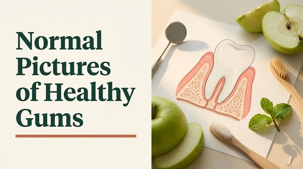
In a healthy mouth, that free gingival margin sits close to the tooth with a tiny, shallow groove called the gingival sulcus between the gum and tooth surface. According to the American Academy of Periodontology (AAP), a healthy sulcus measures between 1 and 3 millimeters in depth. Anything deeper signals that the gum tissue has begun to detach or inflame.
The overall look is tidy. There is no puffiness at the margin. The gums don’t droop over the tooth surface or pull back below it. When you run a clean finger along the gum line, the tissue should feel dense and resistant, not spongy or tender.
Key characteristics of healthy gum tissue:
- Color: pale to medium pink (with natural variation by skin tone; see the section on pigmentation below)
- Texture: stippled or slightly granular, like the surface of an orange peel
- Contour: follows the tooth shape closely, with pointed triangular tissue filling the spaces between teeth (called interdental papillae)
- Attachment: sits firmly at or just above the tooth’s neck with no visible gap or recession
- Bleeding: none when brushing or flossing with normal pressure
Normal Pictures of Healthy Gums: A Visual Reference Guide
When reviewing normal pictures of healthy gums, the most reliable way to interpret what you’re seeing is to know the four visual markers that trained dental professionals use to assess gingival health.
Think of your gum tissue the way a chef evaluates fresh herbs. Fresh basil is vibrant, firm at the stem, consistent in color, and releases no liquid when pressed. Wilted basil is darker, limp, slightly translucent, and bruises at the slightest pressure. Gum tissue works on the same basic principle: healthy tissue is dense and consistent, while inflamed tissue shows its distress through color change, swelling, and fragility.
According to clinical standards published in the Journal of Periodontology, dental professionals assess gingival health using four primary parameters:
| Assessment Marker | Healthy Appearance | Unhealthy Appearance |
|---|---|---|
| Color | Pale to medium pink; uniform | Red, magenta, or deep red |
| Texture | Stippled, firm, orange-peel-like | Smooth, shiny, swollen |
| Contour | Tight knife-edge margin at tooth neck | Rolled, puffy, or blunted margin |
| Bleeding | None on probing or brushing | Bleeds easily with light contact |
Each of these markers tells a story. Color shift toward red indicates increased blood flow from inflammation. Loss of stippling signals swelling beneath the surface. A rolled margin means the tissue can no longer hold its proper shape against the tooth. These are visible changes, and they consistently appear before pain does.
People with naturally darker gingival pigmentation may see a base color that reads as brown or tan rather than pink. That is completely normal and is addressed in detail in the skin tone section below. The inflammatory markers (swelling, shiny texture, bleeding) apply regardless of natural pigmentation color.
What Color Are Healthy Gums
Healthy gums in lighter-skinned individuals are typically described as coral pink or pale salmon pink, but gum color exists on a wide natural spectrum and varies considerably with genetic background and skin tone.
The pink color in gum tissue comes from a combination of blood supply through the underlying capillaries and the translucency of the oral epithelium (the outer tissue layer). In individuals with less melanin in their oral tissues, the blood beneath the surface shows through more clearly, producing that characteristic pink tone.
Gum color by general category:
| Natural Gum Color | Who Typically Shows This | Notes |
|---|---|---|
| Pale pink to coral | Lighter skin tones | Most common reference standard in dental literature |
| Salmon or peachy pink | Medium skin tones | Normal, no cause for concern |
| Brown or tan patches | Darker skin tones, any ethnicity | Called physiologic pigmentation; entirely normal |
| Uniformly brown or dark | Common in individuals of African, South Asian, or Mediterranean descent | Normal variant; consistent pattern is key |
| Pale or white patches | Any skin tone | Requires dental evaluation; not a normal color |
| Bright red or magenta | Any skin tone | Indicates active inflammation regardless of baseline |
The National Institute of Dental and Craniofacial Research (NIDCR) emphasizes that gum color assessment must always account for an individual’s natural baseline. A person whose gums have always been brown-pigmented should look for changes from their personal normal, not comparison against a pink standard that doesn’t apply to them.
For anyone uncertain about whether their gum color is within a healthy range, a dental professional can assess gingival health using probing depth and bleeding on probing, which are objective measurements that don’t depend on color alone.
Key Takeaway: Healthy gums aren’t one specific shade of pink. What matters is firmness, no bleeding, no swelling, a tight fit around each tooth, and no departure from your personal color baseline.
Healthy Gum Texture and What It Tells You
The surface texture of healthy gum tissue has a finely pebbled, slightly rough appearance called stippling, which looks similar to the dimpled surface of an orange peel when examined closely.
Stippling is caused by the attachment of collagen fiber bundles between the epithelial tissue and the underlying connective tissue. Those attachment points create small indentations visible on the surface. The presence of stippling is one of the clearest visible signs that gum tissue is healthy and properly structured.
When gum tissue becomes inflamed, one of the earliest changes is the loss of stippling. The tissue swells with fluid, stretching the surface smooth and giving it a glossy or shiny appearance. If you notice your gum surface looks unusually smooth or reflective compared to how it normally looks, that shift in texture is worth noting and discussing with a dental provider.
What gum texture tells you at a glance:
- Stippled, slightly rough surface: healthy attached gingiva, collagen structure intact
- Smooth, shiny surface: early to moderate inflammation; tissue is edematous (swollen with fluid)
- Granular or cobblestone texture: can indicate chronic inflammation or certain systemic conditions; warrants professional evaluation
- Fibrotic, dense, pale texture: sometimes seen after prolonged inflammation resolves, or as a medication side effect (a condition called gingival hyperplasia, associated with certain blood pressure medications, anticonvulsants, and immunosuppressants)
People taking medications in those drug categories should specifically ask their dentist to assess gum texture changes, as gingival hyperplasia can be subtle in early stages and is not always accompanied by bleeding or pain.
Signs of Healthy Gums: What to Check at Home
Healthy gums show five consistent signs that you can assess yourself with a mirror, good lighting, and clean hands.
A straightforward way to think about a gum health self-check: you’re looking for the same qualities a produce buyer looks for in fresh lettuce. Tight, crisp, no brown edges, no wilting, no water-logging. Healthy gum tissue has that same density and precision.
Five-point home assessment:
- Color check. Look at your gum line in bright natural light. The tissue should be uniform in color across the same areas. Patchy redness at specific teeth is more meaningful than overall uniform pigmentation.
- Bleeding check. Brush and floss normally. Healthy gums do not bleed with standard-pressure brushing or flossing. Occasional minor bleeding when first starting a flossing habit after a long gap is different from consistent bleeding at the same spots.
- Texture check. Run a clean fingertip lightly across the attached gum tissue above your upper teeth. It should feel firm and slightly resistant, not soft, spongy, or tender.
- Contour check. Look at the triangular points of gum tissue between each tooth (the interdental papillae). In a healthy mouth, these points fill the space between teeth completely with no black triangles or gaps.
- Position check. The gum line should sit just at the tooth’s neck. Gum tissue that appears to be pulling away from the tooth, exposing more of the tooth root, indicates recession, which requires professional evaluation.
According to the American Dental Association (ADA), adults should perform a basic gum health self-check monthly in addition to scheduling professional cleanings and examinations at least twice yearly.
Pictures of Healthy Gums vs Unhealthy Gums
A side-by-side comparison of healthy versus unhealthy gum tissue shows immediate, visible differences in color, surface quality, contour, and the way tissue sits against each tooth.
The contrast becomes clearest at the gum margin. In healthy tissue, the margin is thin and knife-edge sharp where it meets the tooth. In inflamed tissue, the margin is rounded, thickened, and often rolls slightly away from the tooth surface, creating a small visible gap. That gap is where bacterial biofilm accumulates and the disease process deepens.
| Feature | Healthy Gums | Unhealthy Gums |
|---|---|---|
| Color | Pale to medium pink (or natural pigment) | Red, magenta, or bluish-red |
| Surface texture | Stippled, firm, matte | Smooth, shiny, glossy |
| Gum margin | Thin, knife-edge, tight to tooth | Swollen, rounded, pulled away |
| Interdental papillae | Fill space between teeth completely | Blunted, missing, or swollen |
| Bleeding | None | Bleeds easily with brushing |
| Pain | None | May be tender or painful |
| Odor | None beyond normal breath variation | Persistent bad breath possible |
The CDC notes that the earliest stage of gum disease, gingivitis, affects an estimated 47.2% of American adults over 30. Gingivitis is entirely reversible with proper care at this stage precisely because the inflammation has not yet destroyed the bone or connective tissue beneath the gum. Catching the visible changes in this comparison chart early is what makes reversal possible.
People who smoke or use tobacco products may have gum tissue that appears paler than expected even when inflamed, because nicotine restricts the blood vessels that normally cause redness. This masking effect means tobacco users cannot rely on color change alone as an early warning sign and benefit from more frequent professional monitoring.
Gingivitis vs Healthy Gums: How to Tell the Difference
Gingivitis is the earliest and fully reversible stage of gum disease, characterized by gum tissue that bleeds, appears red or swollen, and has lost its normal stippled texture, while healthy gums show none of these signs.
The word gingivitis comes from the Latin gingiva (gum) and the suffix -itis (inflammation). It is caused by bacterial plaque accumulating at the gum line. Plaque is a sticky film of bacteria and their byproducts that forms continuously on teeth. When it’s not removed through brushing and flossing, it triggers an immune response in the gum tissue.
Gingivitis vs. healthy gums at a glance:
| Sign | Healthy Gums | Gingivitis |
|---|---|---|
| Bleeding | None | Bleeds when brushing or flossing |
| Color | Pink or natural pigment | Red or bright red at the margin |
| Swelling | None | Noticeable puffiness, especially at margin |
| Stippling | Present | Lost; surface appears smooth and shiny |
| Pain | None | May be tender; often painless |
| Reversibility | N/A | Fully reversible with proper care |
The key clinical distinction is that in gingivitis, the bone and connective tissue supporting the teeth remain intact. The disease has not yet moved below the gum line into the structures that hold teeth in place. According to the AAP, gingivitis can be fully resolved with professional cleaning combined with consistent daily plaque removal at home.
People with diabetes, those who are pregnant, individuals taking certain medications (including some anticonvulsants, calcium channel blockers, and immunosuppressants), and people who smoke are at higher risk of developing gingivitis and should discuss increased monitoring frequency with their dental provider, as these conditions alter the gum tissue’s normal inflammatory response.
Key Takeaway: Gingivitis is the point where the trajectory is still fully in your control. Red gums that bleed during brushing are a signal, not a sentence, and removing plaque consistently reverses the inflammation before it progresses to structural damage.
Gum Disease vs Healthy Gums: The Full Comparison
Gum disease, clinically known as periodontitis, goes beyond gingivitis by destroying the bone and connective tissue that anchor teeth in place, producing changes that are not reversible in the way gingivitis is.
The progression from healthy gum tissue to periodontitis follows a predictable sequence. Plaque that is not removed at the gum line hardens into tartar (also called dental calculus), which cannot be removed by brushing alone. Tartar provides a rough surface for more plaque accumulation and drives deeper inflammation. The immune response begins breaking down the periodontal ligament and alveolar bone beneath the gum.
Stages of gum health from healthy to advanced disease:
| Stage | Clinical Name | Key Signs | Reversibility |
|---|---|---|---|
| Stage 1 | Healthy gingiva | No bleeding, firm, pink/natural, stippled | N/A |
| Stage 2 | Gingivitis | Bleeding, redness, swelling; no bone loss | Fully reversible |
| Stage 3 | Mild periodontitis | Pocket depths 4-5mm, early bone loss on X-ray | Manageable, not reversible |
| Stage 4 | Moderate periodontitis | Pockets 5-7mm, visible recession, tooth sensitivity | Managed, not reversible |
| Stage 5 | Severe periodontitis | Pockets over 7mm, significant bone loss, tooth loosening | Requires specialist intervention |
The NIDCR reports that severe periodontitis affects approximately 8.5% of American adults. At this stage, the gum tissue itself may recede significantly, exposing root surfaces. The gum line appears lower than normal, and teeth may appear longer than they used to. Some people describe a “long tooth” look that wasn’t present before.
Visually, periodontitis-affected gums often show a complex picture: some areas may look less actively red than gingivitis but show clear recession and spacing between teeth that wasn’t there before. This is why assessing gum health by color alone is insufficient.
What Do Healthy Molars Look Like
Healthy gums around the molars (the large, flat-crowned teeth at the back of the mouth) should look identical in color and texture to gum tissue at the front, but the anatomy is more complex and harder to self-examine.
Molars sit in the back of the mouth where visibility is limited, making them a common site for undetected gum disease. The gum tissue between molars (the interdental papillae in those spaces) tends to be broader and wider than the slim pointed papillae visible between front teeth. That broader contact area makes it easier for food and plaque to accumulate undetected.
What healthy gum tissue around molars shows:
- The same pale to medium pink color (or natural pigment) as front gum tissue
- No puffiness or swelling around the molar gum line
- Papillae that fill the space between the molars fully, without gaps
- No exposed root surfaces below the gum margin
- No dark reddish discoloration that differs from the surrounding tissue
Reaching molar gum tissue requires specific flossing techniques and, ideally, an interdental brush (a small brush that fits between teeth) sized for the wider molar contact points. The ADA recommends cleaning between teeth at least once daily, specifically noting that the gum health impact of interdental cleaning is strongest when done consistently.
People with wisdom teeth partially erupted through the gum tissue should be aware that the flap of gum tissue overlying a partially erupted molar (called the operculum) can trap bacteria and becomes a site of infection called pericoronitis. Healthy tissue around fully erupted molars has no overlying gum flap.
Healthy Gum Line Appearance
The healthy gum line follows a consistent, scalloped curve along the jaw, sitting at the same height relative to the crown of each tooth and creating a smooth visual rhythm across the mouth.
Imagine the gum line as the horizon line in a photograph. A healthy horizon is smooth and continuous, with predictable rises and dips following the tooth anatomy. An unhealthy one shows irregular breaks, sudden drops, and patches where the line has retreated or swollen unevenly.
In a healthy adult mouth:
- Upper front teeth: the gum line peaks at the center of each tooth, creating a gentle scallop shape
- Lower front teeth: the gum line is slightly flatter, following the shorter crowns of the lower incisors
- Canine teeth: the gum line tends to peak slightly higher at the canine tips, which is normal anatomy
- Premolars and molars: the gum line is broader, less scalloped, and more straight across
A gum line that has dropped unevenly at specific teeth, exposing the yellower root surface below the original margin, indicates gum recession at those sites. Recession can result from gum disease, overly aggressive brushing with a hard-bristled toothbrush, orthodontic movement, or in some cases genetic predisposition to thin gum tissue.
According to a clinical review published in the Journal of Clinical Periodontology, recession affecting more than 2 millimeters of root surface at any single tooth site warrants professional assessment, as root surfaces exposed to the oral environment are significantly more vulnerable to decay than crown enamel.
Key Takeaway: The gum line is a map. An uneven map tells a story about where disease, recession, or trauma has occurred, and a consistent, scalloped line that sits properly at each tooth neck is one of the clearest signs a mouth is healthy.
Healthy Teeth and Gums: What the Whole Mouth Should Look Like
A healthy mouth combines firm, properly colored gum tissue with tooth surfaces that are intact, and the relationship between gum tissue and tooth at every site looks consistent and proportional.
The proportion between tooth crown and visible gum tissue is called the gingival-tooth ratio. In a healthy adult mouth, approximately one-third to one-quarter of the total tooth length is normally visible as the crown, with the gum covering the remaining root. When that proportion looks off, either because the gums have receded (showing too much tooth) or because the gums overgrow the crown (showing too little), something is affecting normal anatomy.
What a full healthy mouth assessment covers:
| Feature | Healthy Appearance |
|---|---|
| Gum color | Uniform, pale to medium pink or natural pigment |
| Gum texture | Stippled, firm, not shiny |
| Gum margin | Tight, no gaps or recession |
| Interdental papillae | Fill all spaces between teeth |
| Tooth surfaces | White to off-white enamel, no dark discoloration at gum line |
| Saliva | Clear and thin; mouth feels moist |
| Breath | Neutral; no persistent odor |
| Tooth stability | No mobility or looseness when touched |
Tooth color itself varies naturally. Enamel ranges from bright white to a natural ivory or slightly yellow tone, and that variation is entirely normal. What is not normal is a dark brown or black line at the gum margin of any tooth, which can indicate secondary decay at an old restoration or calculus buildup requiring professional removal.
People who wear orthodontic brackets should pay particular attention to the gum tissue directly beneath and around each bracket, as brackets create areas where plaque accumulates out of easy reach and gingivitis around brackets is one of the most common side effects of braces treatment.
Gum Color Variation by Skin Tone: What Is Normal for You
People with darker skin tones frequently have gum tissue that is brown, tan, or has patches of darker pigmentation, which is a completely normal anatomical feature caused by melanin deposition in the gingival tissue.
This pigmentation is called physiologic pigmentation or racial pigmentation in clinical literature. It is not a sign of disease. It is not harmful. It does not require treatment. The NIDCR explicitly recognizes this natural variation and notes that dental assessment of gingival health in individuals with pigmented gum tissue relies on changes in texture, contour, and bleeding on probing rather than color alone.
Gum pigmentation patterns by background:
| Population Group | Common Pigmentation Pattern | Notes |
|---|---|---|
| Northern European descent | Uniformly pale or coral pink | Less melanin in gingival tissue |
| Mediterranean descent | Pink with possible brown-gray patches | Mixed pigmentation common |
| South Asian descent | Brown or dark tan, often uniform | Physiologic; entirely normal |
| East Asian descent | Light pink to mild brownish patches | Moderate variation; usually light |
| African descent | Dark brown to nearly black tissue possible | Deeply pigmented; completely normal |
| Latin American descent | Wide range; pink to significant brown | Reflects mixed ancestry patterns |
The warning sign in pigmented gum tissue is not the base color itself but a change from the person’s established baseline. Suddenly developing new dark patches where the gum was previously lighter, or patches that bleed when touched, should be evaluated by a dental professional.
One specific condition worth knowing: a blue-black line running precisely along the gum margin, especially near old metal restorations, may indicate amalgam tattoo (harmless) or, less commonly, an early melanotic lesion that warrants examination. Consistent uniform pigmentation is different from newly appearing localized spots.
Healthy Gums After Deep Cleaning: What to Expect
After a professional scaling and root planing procedure (commonly called a deep cleaning), healthy-looking gum tissue begins to show within 2 to 4 weeks as inflammation resolves and the tissue reattaches to the cleaned root surfaces.
Scaling and root planing is the primary non-surgical treatment for periodontitis. It involves removing calculus and bacterial biofilm from root surfaces below the gum line, which the gum tissue then begins to heal against. The immediate post-treatment appearance is often alarming to patients. The gums may look more receded, redder, or slightly swollen right after the procedure. This is normal and expected.
Recovery timeline after deep cleaning:
| Timeframe | Expected Gum Appearance |
|---|---|
| Day 1 to 3 | Tenderness, mild swelling, possible minor bleeding |
| Day 4 to 7 | Swelling begins to reduce; tissue looks less red |
| Week 2 | Redness decreasing; gum margin starts to tighten |
| Week 3 to 4 | Stippling returns; tissue becomes firmer |
| Month 2 to 3 | Full healing; dentist reassesses pocket depths |
According to research published in the Journal of Periodontology, most patients show measurable reduction in pocket depth and improvement in gingival health indices within 6 to 8 weeks of scaling and root planing when combined with consistent home care.
The gum line may appear slightly lower than before the procedure because swollen tissue that was covering the root now sits in its proper, healthy position. This can make teeth look slightly longer. This is not recession; it is the result of inflammation resolving. Your dental provider is the most reliable person to confirm whether what you’re seeing reflects healthy healing or a concern, given that individual healing rates vary based on the extent of disease, systemic health, and home care consistency.
Key Takeaway: Post-deep-cleaning gum tissue looks worse before it looks better. Redness and apparent recession in the first week are normal parts of healing, not evidence the treatment failed.
Healthy Gums During Pregnancy and Hormonal Changes
Pregnancy and other hormonal shifts can cause gum tissue to become more sensitive, more prone to bleeding, and more prone to swelling even with healthy oral hygiene habits. This is a well-documented physiological response, not a reflection of poor dental care.
The condition is called pregnancy gingivitis, and it affects an estimated 60 to 75% of pregnant people according to data reviewed in the Journal of Clinical Periodontology. Rising levels of estrogen and progesterone increase blood flow to gum tissue and alter the tissue’s inflammatory response, making it react more strongly to the same amount of plaque that would not have caused visible inflammation before pregnancy.
How hormonal changes affect gum appearance:
- Gum tissue appears redder and more vascular during the second trimester
- Bleeding during brushing increases even with careful technique
- Some people develop a localized overgrowth of gum tissue at a single site called a pregnancy epulis (also called a pyogenic granuloma), which appears as a small, red, smooth bump at the gum margin
- Tissue swelling can make gum pockets temporarily deeper, creating conditions that favor plaque retention
The AAP recommends that pregnant individuals maintain their regular dental cleaning schedule and inform their dental provider of the pregnancy, as professional cleaning during pregnancy is safe and helps manage the increased inflammatory risk.
Puberty, menopause, and the use of oral contraceptives can produce similar, though generally milder, changes in gum tissue. Anyone who notices a change in their gum appearance or bleeding pattern that coincides with a hormonal shift should discuss this connection with their dental provider. The underlying cause shapes the treatment approach.
How to Get and Maintain Healthy Gums
Healthy gums are maintained through a combination of mechanical plaque removal (brushing and flossing), regular professional cleaning, and several nutrition-based factors that support the integrity of gingival tissue.
The mechanics of plaque removal matter more than the product used. A soft-bristled toothbrush used with gentle, circular motions at a 45-degree angle to the gum line removes plaque from both the tooth surface and the gingival margin without traumatizing the tissue. The ADA recommends brushing for two full minutes, twice daily.
Daily habits that support gingival health:
- Brush for two minutes, twice daily, with a soft-bristled brush at a 45-degree angle to the gum line
- Clean between teeth at least once daily using floss, interdental brushes, or a water flosser (all are effective when used consistently)
- Replace a toothbrush or brush head every 3 months or sooner if bristles are visibly worn
- Avoid tobacco in any form; tobacco restricts circulation to gum tissue and masks early inflammation
- Drink water rather than acidic or sugar-sweetened beverages throughout the day to support a balanced oral microbiome
- Schedule professional cleaning and examination at least twice yearly
Nutrition that supports gum tissue:
Vitamin C (ascorbic acid) is directly involved in collagen synthesis in gingival connective tissue. The NIDCR notes that severe vitamin C deficiency produces a condition called scurvy, which includes gingival bleeding and tissue breakdown as primary symptoms. While outright scurvy is rare, inadequate vitamin C intake has been associated with poorer periodontal outcomes in observational studies.
Vitamin D plays a role in periodontal immune regulation. A 2020 review in the Journal of Periodontology found that individuals with lower serum vitamin D levels showed higher rates of periodontal disease than those with adequate levels. Good dietary sources of vitamin D include fortified dairy products, salmon, tuna, egg yolks, and fortified plant-based milks.
People who wear dental implants need to note that the gum tissue around an implant (called peri-implant mucosa) requires the same maintenance as natural tooth gingiva and is susceptible to its own form of inflammation called peri-implantitis. Standard brushing and daily cleaning around implants is essential, and some implant geometries benefit from specific brush types recommended by the restoring dentist.
Frequently Asked Questions About Healthy Gums
What is the normal color of healthy gums?
Healthy gums are normally pale to medium pink in lighter-skinned individuals, but natural gum color varies from coral pink to brown or dark tan depending on an individual’s natural melanin levels.
The key is consistency within your own mouth. Uniform color that matches your personal baseline is a healthy sign.
Sudden color changes, especially redness, dark patches that appear where they weren’t before, or white patches at any skin tone, warrant dental evaluation.
How do I know if my gums are healthy or not?
Healthy gums don’t bleed when you brush or floss with normal pressure, feel firm and slightly rough in texture, and sit tightly against each tooth without gaps or recession.
If your gums bleed consistently, look swollen or shiny, or if you notice the gum line dropping away from your teeth, these are signs to discuss with a dental professional.
A dental provider can confirm gum health using a probe that measures the depth of the pocket between gum and tooth, which is more precise than visual assessment alone.
What do healthy gums look like after a deep cleaning?
In the first one to two weeks after a deep cleaning, healthy healing gums may appear slightly more receded and mildly red as swelling resolves, which is a normal part of the process.
By weeks three to four, stippling returns, the tissue firms up, and the gum line settles into its proper position.
Full tissue assessment by your dental provider typically happens at a follow-up appointment six to eight weeks after the procedure.
Can dark or brown gums be healthy?
Yes, dark or brown gum tissue is completely healthy when it reflects natural melanin pigmentation, which is extremely common in individuals of African, South Asian, Mediterranean, and mixed ethnic backgrounds.
This natural coloring is called physiologic pigmentation and requires no treatment.
The distinction to watch for is a sudden change from your personal baseline color, particularly newly appearing dark spots or lesions that bleed on contact.
What does a healthy gum line look like around teeth?
A healthy gum line follows a smooth, scalloped curve that sits at the base of the tooth crown, with pointed triangular tissue filling the spaces between teeth completely.
It should be consistent in height across the mouth, with no areas where the gum has visibly pulled back from the tooth surface.
Uneven gum line height, exposed root surfaces that appear yellower than the crown, or black triangles visible between teeth indicate recession or tissue loss that a dentist should evaluate.
Closing
Healthy gum tissue is specific and recognizable once you know what to look for. Firm, properly contoured, non-bleeding, and consistent with your personal color baseline are the markers that actually matter. The comparison tables in this guide give you a real framework, not just vague reassurance.
The most useful thing you can do today is spend two minutes in good light and actually look at your gums before your next dental appointment. Know your baseline. That way, any change becomes obvious rather than something you’ve lived with long enough to stop noticing.
Regular professional assessment remains the most reliable check. If anything in this guide prompted a specific concern, bring it to your next dental visit with the specific details of what you observed and where.