
Healthy gums are pale pink, firm, and fit tightly around each tooth without gaps, puffiness, or bleeding. If yours match that description, your gum tissue is doing its job properly.
Gum health matters more than most people realize. According to the Centers for Disease Control and Prevention (CDC), nearly 47.2% of adults aged 30 and over in the United States have some form of periodontal (gum) disease. Many of them had no idea anything was wrong until the damage was already underway.
This guide covers what healthy gum tissue actually looks like, how to read the differences between healthy and diseased tissue, what the science says about diet and gum health, and which foods can genuinely support better gum tissue from the inside out.
What Do Healthy Gums Look Like?
Healthy gums look like pale pink, firm tissue that hugs each tooth cleanly without swelling, redness, or visible separation from the tooth surface.
That description is the clinical baseline used by the American Academy of Periodontology (AAP). The AAP defines periodontal health as the absence of inflammation, bleeding on probing, and bone or attachment loss.
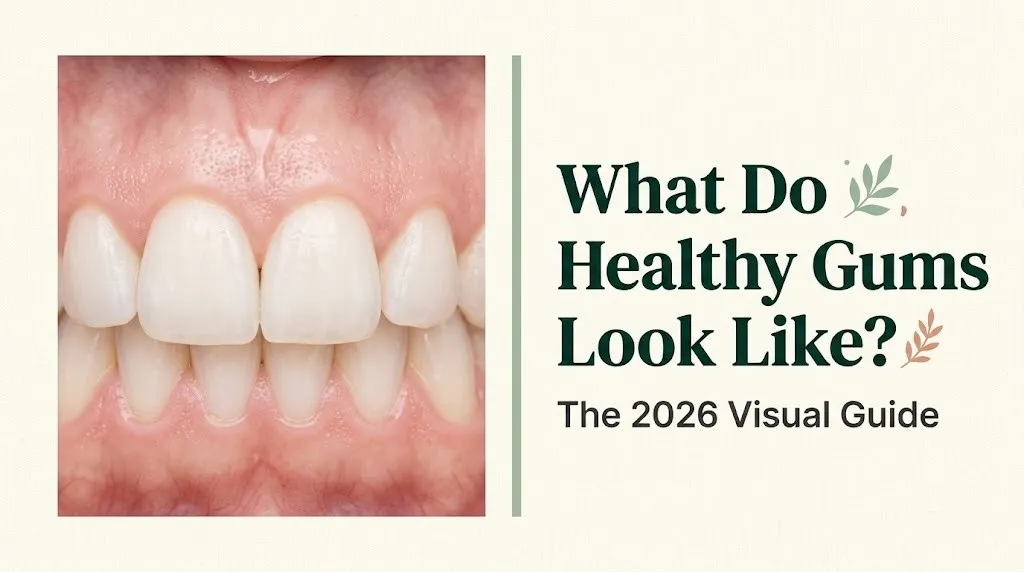
Think of healthy gum tissue like a well-fitted collar around each tooth. It should sit snugly, lie flat, and show no signs of strain. A collar that’s puffed out, red, or pulling away from the shirt signals a problem.
The key visual markers dentists use include:
- Color: Pale pink or coral pink (with natural variation explained in the next section)
- Texture: Firm and slightly stippled (a finely pebbled or orange-peel surface)
- Shape: Knife-edged or pointed between teeth, not rounded or bulging
- Fit: Tight against the tooth surface, no visible gaps or pockets
- Bleeding: None during gentle brushing or flossing
- Odor: No persistent bad odor from the gum tissue itself
The absence of bleeding is one of the most reliable home-observable signs of gum health. According to research published in the Journal of Clinical Periodontology, bleeding on probing is among the earliest measurable clinical indicators of gingival (gum) inflammation.
What Should Healthy Gums Look Like: The Core Visual Checklist
Healthy gums should be firm, pale pink, non-tender, and closely adapted to each tooth with no puffiness, bleeding, or recession visible.
Here is the complete visual checklist that a periodontist uses during a routine exam, translated into what you can actually observe at home with a bathroom mirror and a small flashlight:
| Visual Feature | What Healthy Looks Like | What to Watch For |
|---|---|---|
| Color | Pale pink or coral pink | Red, dark red, or purple hue |
| Texture | Slightly stippled, firm surface | Smooth, shiny, or puffy surface |
| Gum line shape | Pointed peak between teeth | Rounded, swollen, or flattened peaks |
| Fit around tooth | Tight, no visible gap | Gap, pocket, or visible root |
| Bleeding | None when brushing gently | Bleeds with light pressure |
| Swelling | No puffiness | Raised, swollen tissue |
| Pain or tenderness | None at rest or when touched | Pain, soreness, or sensitivity |
None of these markers alone tells the complete story. A dentist or periodontist uses a periodontal probe to measure sulcus depth (the shallow groove where gum meets tooth). A healthy sulcus depth is 1 to 3 millimeters. Depths above 4 millimeters suggest the beginning of a periodontal pocket.
People taking certain medications, including some calcium channel blockers and anticonvulsants, may experience gum overgrowth (gingival hyperplasia) that alters the appearance even without infection. If your gum tissue looks enlarged but not red, mention your full medication list to your dentist.
Normal Healthy Gum Color
Normal healthy gum color ranges from pale pink to coral pink in people with lighter skin tones, and from coral pink to brown or deep brown in people with higher natural melanin levels in their oral tissue.
This is one of the most misunderstood areas in gum health content, and it causes real anxiety. The gingiva (gum tissue) contains melanocytes, the same pigment-producing cells found in skin. People of African, South Asian, East Asian, Mediterranean, and Hispanic descent commonly have naturally darker gum pigmentation. This is a normal anatomical variation. It is not a sign of disease.
The American Dental Association (ADA) and the AAP both acknowledge that healthy gum color varies substantially based on a person’s natural melanin level. A dentist assesses color in context: they look for changes from that individual’s baseline, not against a single universal standard.
What matters most for color assessment:
- Uniformity: Healthy gums show consistent coloring across the tissue. Patchy redness, localized purple spots, or sudden color change against your personal baseline are worth monitoring.
- Redness as a signal: In any skin tone, active red or dark red coloring around the gum margin suggests increased blood flow from inflammation, not normal pigmentation.
- White or gray patches: These are never a normal color variation and should be evaluated by a dentist promptly. White patches can indicate a range of conditions, including oral candidiasis or potentially precancerous lesions.
Quick Tip:
- Take a well-lit photo of your gums every six months to build a personal baseline.
- Compare photos over time for any clear shifts in color, shape, or fit around your teeth.
- Report any sudden color change to your dentist even if there’s no pain.
Key Takeaway: Healthy gums are pale to coral pink, firm, non-bleeding, and tightly fitted around each tooth. But “healthy pink” isn’t one single shade. Your baseline color depends on your natural melanin level, and any change from your own baseline is what actually signals a problem.
Healthy Gum Texture and Firmness
Healthy gum tissue feels firm and slightly resilient when you press it gently, and its surface has a finely textured, almost pebbled appearance called stippling.
Stippling looks like the surface of an orange peel, just at a much smaller scale. It comes from the way collagen fibers attach the outer gum tissue (the attached gingiva) to the underlying bone. This texture is a sign that the tissue is well-organized and structurally sound.
Not everyone shows stippling equally. Children often have smoother gum surfaces that develop more texture with age. Some adults naturally have less visible stippling even with perfectly healthy tissue. The presence of stippling is a positive sign, but its absence alone does not confirm disease.
Texture changes that indicate a problem include:
- Smooth and shiny surface: This is characteristic of inflamed tissue. When inflammation causes the gum tissue to swell, it stretches the surface and erases the normal stippling pattern.
- Spongy or soft feel: Healthy gums feel firm. Tissue that compresses noticeably under light finger pressure has lost structural integrity.
- Raised or cord-like margin: The free gingival margin (the very edge of the gum where it meets the tooth) should be thin and knife-like. A thickened, cord-like margin suggests chronic inflammation.
According to the Journal of Periodontology, stippling loss is one of the early clinical tissue changes associated with gingival inflammation, appearing before significant bleeding or pocketing develops.
Healthy Gum Line: What the Gum-Tooth Border Should Look Like
The healthy gum line follows a smooth, scalloped arc that traces the curve of each tooth, sitting just above the base of the visible tooth crown without exposing any root surface.
The cementoenamel junction (CEJ) is the anatomical border where the enamel (tooth crown) meets the cementum (tooth root). In a healthy mouth, the gum line sits slightly above the CEJ so the root is completely covered. No root exposure is normal. Any visible root surface means the gum has receded from where it should be.
Between each pair of adjacent teeth, the gum fills the space with a pointed peak of tissue called the interdental papilla. In healthy gum tissue, this papilla:
- Fills the entire triangular space between adjacent teeth
- Has a pointed or slightly rounded tip, not a flat or blunted one
- Shows no swelling or redness at its tip
- Does not bleed when flossed correctly
When the interdental papilla is lost or blunted, it creates what dentists call “black triangles” (the dark spaces visible between teeth near the gum line). This is a common sign of gum recession or previous inflammation. It is also a common cosmetic concern following orthodontic treatment.
| Gum Line Feature | Healthy Appearance | Problematic Appearance |
|---|---|---|
| Margin position | Just above the CEJ | Pulled away, exposing root |
| Interdental papilla | Full, pointed, fills tooth gap | Blunted, absent, or swollen |
| Scalloping pattern | Smooth arc, consistent depth | Uneven, jagged, or asymmetric |
| Margin thickness | Thin and knife-edged | Thick, rolled, or cord-like |
Healthy Gums Inside the Mouth: Different Areas Explained
Healthy gum tissue inside the mouth looks consistent across all zones, but the tissue type, thickness, and appearance vary between the gum that hugs the teeth and the softer tissue further back in the mouth.
There are two distinct types of gum tissue in your mouth:
1. Attached gingiva: This is the firm, stippled, pale pink tissue directly around the teeth. It is tightly bound to the underlying alveolar bone (the bone that holds your teeth). This tissue is meant to be immovable and firm.
2. Alveolar mucosa: This is the redder, shinier, and softer tissue further from the teeth, toward the inner cheeks and throat. It is normal for this tissue to look darker and more vascular. Many people mistake the appearance of healthy alveolar mucosa for gum disease because it is naturally redder.
The border between these two tissue types is called the mucogingival junction. In a healthy mouth, this line is clearly visible as a distinct color shift from the pale attached gingiva to the deeper-red alveolar mucosa.
Behind the lower front teeth (the lingual gum), the tissue tends to be slightly thinner and may look slightly paler or more translucent. This is normal. The tissue at the back of the mouth around the molars tends to be thicker.
Quick Tip:
- Use a small dental mirror or the front camera on your phone to check the gum tissue on the tongue side of your lower front teeth, where early recession is often first visible.
- The tissue color shift at the mucogingival junction is normal and not a sign of disease.
Key Takeaway: Not all red tissue in your mouth is gum disease. The alveolar mucosa is naturally darker and softer than attached gingival tissue, and understanding which is which prevents a lot of unnecessary alarm.
Healthy Lower Gums and Bottom Teeth
Healthy lower gums look the same as upper gums in terms of color and firmness, but the tissue tends to be slightly thinner and the gum line often sits closer to the bottom teeth roots because lower front teeth have naturally shallower bone support.
The lower front teeth (mandibular incisors) are among the most common sites for gum recession, not because of disease necessarily, but because the bone on the outer (lip-facing) side of these teeth is naturally thinner. Thin bone means thinner overlying gum tissue, which is more susceptible to recession from aggressive brushing.
Healthy lower gums in this zone should still:
- Cover the full root surface with no root exposure
- Show no redness or swelling at the gum margin
- Have a complete interdental papilla between each lower front tooth
- Produce no pain when touched or flossed
The gum tissue behind the lower front teeth (on the tongue side, called the lingual aspect) is frequently overlooked during self-exams. This tissue should be pale pink and firm, identical to the tissue on the outer side.
The National Institute of Dental and Craniofacial Research (NIDCR) notes that recession on the lower front teeth is one of the most common findings in adults who brush with a hard-bristled toothbrush or use horizontal scrubbing motions. Switching to a soft-bristled brush with a circular technique can halt progression.
For people with lower gum sensitivity or visible recession, a periodontist referral is appropriate to measure whether recession is stable or actively progressing.
Healthy Upper Gums
Healthy upper gums are pale pink, firm, non-swollen, and show a complete, symmetrical gum line across all upper teeth, including the back molars where the gum tissue is naturally thicker.
Upper gum tissue tends to be thicker than lower gum tissue because the upper jaw (maxilla) has denser bone support. This means upper gums are generally more resistant to recession. However, they are not immune to it.
The upper front teeth are a frequent site of inflammation because plaque accumulates along the gum line where the teeth slightly overlap. Redness, swelling, or bleeding specifically at the top center of the gum arch, between the two upper front teeth, often reflects inadequate flossing in that specific spot.
Upper gum health also includes:
- The tissue along the roof of the mouth (palate), which is naturally pale pink to white and slightly ridged near the front. This is normal palatine tissue, not gum disease.
- The gum tissue around upper molars, which should be thick, firm, and show no gaps or pocketing even though these areas are harder to see and clean.
- The gum line at the back of the upper jaw, where the wisdom tooth sites are, if teeth are present. This tissue should fit snugly with no open flaps or pockets.
| Location | Normal Gum Appearance | Common Issue in This Zone |
|---|---|---|
| Upper front teeth | Symmetrical scallop, pink | Inflammation from missed flossing |
| Upper back molars | Thick, firm, no pocketing | Deeper pockets hard to clean |
| Palate tissue | Pale, ridged near front | Often mistaken for abnormal |
| Wisdom tooth site | Snug, no flap or pocket | Pericoronitis if partially erupted |
Key Takeaway: Upper gum tissue is naturally thicker than lower gum tissue, making it more resilient to recession, but the upper front gum line between the two central incisors is often the first place inflammation appears when flossing is skipped.
Healthy Gums vs Unhealthy Gums: Full Comparison
Healthy gums are pale pink, firm, non-bleeding, and tightly fitted to each tooth. Unhealthy gums are red or purple, swollen, soft, prone to bleeding, and may show gaps between the gum and tooth surface.
This is the comparison most people are actually searching for, and no single bullet list does it justice. Here is a detailed side-by-side:
| Feature | Healthy Gums | Unhealthy Gums |
|---|---|---|
| Color | Pale pink to coral pink | Red, bright red, dark red, purple |
| Texture | Firm, slightly stippled (pebbled) | Smooth, shiny, spongy |
| Swelling | No swelling, fits tooth tightly | Puffy, raised, or engorged |
| Bleeding | None with normal brushing/flossing | Bleeds easily, sometimes spontaneously |
| Pain | No pain at rest or when touched | Tender, sore, or sensitive to touch |
| Gum line | Stable, no root exposure | Receding, root surface visible |
| Odor | None specific to gum tissue | Persistent bad breath from tissue |
| Interdental papilla | Full and pointed | Blunted, absent, or ulcerated |
| Sulcus depth | 1 to 3 mm | 4 mm or more (periodontal pocket) |
The progression from healthy to diseased tissue typically follows a recognizable pattern. Gingivitis is the earliest stage, where inflammation is limited to the gum tissue itself. At this stage, the damage is fully reversible with proper cleaning. Periodontitis represents the advanced stage, where infection has spread below the gum line and begun destroying the bone and ligaments that hold teeth in place. This stage causes permanent bone loss.
According to the CDC, roughly 70.1% of adults aged 65 and over have periodontitis. Early-stage gingivitis affects a much broader portion of the population, often without any pain.
Healthy vs Inflamed Gums: Early Warning Signs
Healthy gums produce no bleeding and no swelling. Inflamed gums begin with a slight change in color from pale pink to a brighter or darker pink-red, followed quickly by swelling and bleeding when touched.
The earliest stage of gum inflammation is called gingival inflammation or gingivitis, and it has a specific clinical definition: tissue that bleeds on gentle probing, even if it still looks nearly normal to the naked eye.
Home-observable early warning signs that your gums have shifted from healthy to inflamed:
- Bleeding when you floss, even gently
- Bleeding when brushing, especially along the gum line
- A slight puffiness at the gum margin that wasn’t there before
- Gums that look slightly brighter or more vivid pink than usual
- A persistent bad taste or mild odor that brushing alone doesn’t resolve
- Mild tenderness when you press the gum tissue with a finger
The transition from gingivitis to periodontitis is not instant. The American Academy of Periodontology (AAP) explains that gingivitis can remain stable at the gingival tissue level for years if oral hygiene improves. The risk is that pain is often absent, which leads people to ignore early warning signs.
One analogy that makes this real: think of inflamed gums like a slow water leak behind a wall. You may not see structural damage for months, but the damage is accumulating the entire time. The moment you notice the stain on the wall (bleeding, recession, loose teeth), serious work has already been done.
If bleeding persists beyond two weeks of consistent twice-daily brushing and daily flossing, scheduling an exam with a dentist or periodontist is the appropriate next step.
Healthy Gums vs Smokers Gums
Healthy non-smoking gums bleed predictably when inflamed and show normal pink color. Smokers’ gums often appear falsely healthy because nicotine reduces blood flow to gum tissue, suppressing the redness and bleeding that would normally signal a problem.
This is one of the most dangerous deceptions in oral health. Tobacco use (cigarettes, cigars, pipe tobacco, and smokeless tobacco) has a vasoconstricting effect on gum tissue, meaning it narrows the blood vessels that supply the gingiva. The result is that gum tissue in smokers often looks pale and does not bleed as readily, which can mislead both the person and a less experienced clinician into thinking the gums are healthy.
The AAP reports that smokers have a significantly higher risk of periodontal disease than non-smokers, and that the disease often progresses further before being detected because the standard clinical warning signs (redness, bleeding) are masked.
Specific differences between healthy non-smoker gums and smoker gums:
| Feature | Healthy Non-Smoker Gums | Typical Smoker Gums |
|---|---|---|
| Color | Pale pink | Pale, grayish, or mottled |
| Bleeding | Bleeds if inflamed | Reduced bleeding despite inflammation |
| Tissue quality | Firm and well-vascularized | Thickened, fibrotic, less resilient |
| Gum line | Stable | Higher recession risk long-term |
| Healing capacity | Normal post-procedure healing | Slowed healing after procedures |
| Disease masking | Inflammation visible as redness | Inflammation hidden by vasoconstriction |
Quitting tobacco use allows gum tissue vascularity to return. Research published in the Journal of Clinical Periodontology has shown measurable improvement in gingival health within eight weeks of smoking cessation, including normalized bleeding response and improved tissue tone.
Key Takeaway: Smokers should not interpret the absence of bleeding gums as a sign of health. Tobacco actively suppresses the visible signals of gum disease, making regular professional periodontal exams especially important for anyone who smokes or has smoked.
What Does Gum Recession Look Like vs Healthy Gums
Healthy gums cover the full root surface of every tooth. Receding gums have pulled away from the tooth, exposing the root, making the tooth appear longer, and often creating sensitivity to temperature or sweet foods.
Gum recession (gingival recession) is the apical (downward or inward) migration of the free gingival margin away from the crown and toward the root. Once gum tissue has receded, it does not regenerate on its own. Prevention and slowing progression are the priorities.
Signs that gums are receding rather than simply sitting at a healthy position:
- Teeth look noticeably longer than they used to
- A yellowish or darker surface is visible at the base of some teeth (this is root cementum, not enamel)
- Sensitivity to cold drinks or cold air at specific teeth
- A visible notch or groove at the gum line of affected teeth
- Spaces between teeth near the gum line that were not there before
Common causes of recession include:
- Aggressive brushing with a medium or hard-bristled toothbrush
- Chronic gum disease (periodontitis) causing bone and tissue loss
- Orthodontic tooth movement outside the natural bone envelope
- Genetic predisposition to thin gum tissue (thin gingival biotype)
- Lip or tongue piercings that mechanically abrade gum tissue
A periodontist can measure whether recession is stable or progressing by comparing sequential measurements over time. Gum grafting procedures can surgically restore coverage of exposed root surfaces. A connective tissue graft (using tissue from the palate) is the most common technique. Individual candidacy depends on recession extent, tissue quality, and other health factors, which a periodontist should evaluate directly.
Food for Healthy Gums: What to Eat
The best foods for healthy gums are those that supply vitamin C (ascorbic acid), vitamin D (cholecalciferol), calcium, folate (vitamin B9), and coenzyme Q10 (CoQ10), all of which directly support gum connective tissue, immune response, and periodontal bone structure.
Gum tissue is primarily made of collagen, a structural protein that requires ascorbic acid (vitamin C) for synthesis. Without adequate vitamin C, collagen production slows, gum tissue weakens, and bleeding becomes more likely. This is why the classic sign of scurvy (severe vitamin C deficiency) is bleeding, spongy gums. You don’t need to be severely deficient to notice an effect. Chronically low vitamin C intake creates subtle gum tissue vulnerability over time.
Research from Harvard T.H. Chan School of Public Health and related studies on diet-periodontal health relationships has consistently associated higher vegetable and fruit intake with lower rates of periodontal disease.
| Nutrient | Function for Gum Health | Best Food Sources |
|---|---|---|
| Vitamin C (ascorbic acid) | Collagen synthesis, immune defense | Bell peppers, kiwi, citrus, strawberries |
| Vitamin D (cholecalciferol) | Anti-inflammatory, supports bone density | Fatty fish (salmon, mackerel), fortified milk |
| Calcium | Alveolar bone strength | Dairy, fortified plant milk, broccoli, kale |
| Folate (vitamin B9) | Reduces gum inflammation markers | Spinach, lentils, chickpeas, asparagus |
| CoQ10 | Gum tissue energy metabolism | Beef, sardines, spinach, cauliflower |
| Phosphorus | Bone mineral density support | Eggs, fish, legumes, nuts |
People on blood-thinning medications (such as warfarin/Coumadin) should maintain consistent rather than dramatically increased intake of high-vitamin K leafy greens. Sudden large increases can affect medication dosing. Discuss any significant dietary changes with the prescribing provider, particularly when increasing leafy green consumption substantially.
How to Maintain Healthy-Looking Gums
Maintaining healthy-looking gums requires consistent mechanical plaque removal, the right brushing technique, and specific dietary habits that support gum tissue from within.
Dental plaque is a biofilm: a structured community of oral bacteria that adheres to tooth surfaces and the gum margin. Within 24 hours of removal, plaque begins reforming. Within 48 to 72 hours without disruption, plaque can trigger the first stages of gingival inflammation. This is why daily consistency in oral hygiene matters more than occasional intense cleaning sessions.
The ADA recommends:
- Brushing for 2 minutes, twice daily, with a soft-bristled toothbrush and fluoride toothpaste
- Cleaning between teeth once daily with floss or an interdental brush
- Using an antimicrobial mouth rinse if recommended by a dentist
- Professional cleanings every 6 months, or more frequently for those with a history of gum disease
Beyond daily hygiene:
- Replace your toothbrush or brush head every 3 months, or sooner if bristles are frayed
- Use a technique that angles the brush at 45 degrees to the gum line, directing bristle tips into the sulcus
- Avoid smoking and limit alcohol, both of which impair gum tissue healing and immune response
- Manage conditions like type 2 diabetes, which the AAP has identified as bidirectionally linked to periodontal disease severity. Poorly controlled blood sugar worsens gum inflammation, and advanced gum disease can make blood sugar harder to control.
People who have completed gum disease treatment (scaling and root planing, or periodontal surgery) should follow the periodontal maintenance schedule their treating clinician establishes. This is typically every 3 to 4 months rather than the standard 6-month interval, and the specific interval should be confirmed with the treating periodontist based on individual disease history and tissue response.
Key Takeaway: Healthy gums are maintained primarily through daily plaque disruption. No mouthwash, food, or supplement replaces the mechanical removal of biofilm from the tooth-gum margin every single day.
Frequently Asked Questions About Healthy Gums
What color should healthy gums be?
Healthy gums should be pale pink to coral pink in people with lower melanin levels in their oral tissue.
In people with higher natural melanin levels, healthy gum color ranges from coral to medium or dark brown, and this is a normal anatomical variation, not a sign of disease.
The key indicator is consistency across your tissue, and any change from your personal baseline color warrants attention.
Is it normal for gums to be dark or have dark spots?
Dark gum pigmentation is completely normal in people with naturally higher melanin levels, including many people of African, South Asian, East Asian, Mediterranean, and Hispanic descent.
The ADA and AAP both recognize natural gingival pigmentation as a normal anatomical variation with no clinical significance.
However, a sudden dark spot that was not there before, especially one that is isolated, raised, or growing, should be evaluated by a dentist to rule out other causes.
What do unhealthy gums feel like?
Unhealthy gums may feel tender, sore, or sensitive to pressure even when not being brushed or touched.
Spongy or soft texture when you press gum tissue with a finger is a common sign of inflammation.
Pain is not always present in gum disease, which is why visual and professional assessment matters even when you feel nothing unusual.
Can your gums go back to being healthy after gum disease?
Gingivitis (the earliest stage of gum disease, limited to gum tissue only) is fully reversible with consistent daily plaque removal and professional cleaning.
Periodontitis (advanced gum disease involving bone loss) is not reversible, but it is controllable with professional treatment and long-term maintenance.
Once bone supporting the teeth is lost, it does not regenerate without surgical intervention, which is why early detection and consistent hygiene make a measurable difference.
What foods are best for keeping gums healthy?
The best foods for gum health are those high in vitamin C (ascorbic acid) (bell peppers, kiwi, citrus fruits), vitamin D (cholecalciferol) (fatty fish, fortified dairy), and calcium (dairy products, fortified plant milks, leafy greens).
Folate-rich foods like spinach and lentils have been associated with reduced gum inflammation markers in research examining diet-periodontal relationships.
No food replaces mechanical plaque removal, but consistent intake of these nutrients supports the structural integrity of gum connective tissue.
Closing
Healthy gums give you clear, readable signals when something is off. The color shifts first. Then texture. Then bleeding. Then recession. Each stage is measurable and, in the early stages, reversible.
Start with a two-minute mirror check using good lighting. Look at color, shape, and fit. Note your personal baseline. If something has changed in the last few weeks, that’s the signal worth acting on.
Book a professional exam if you have any bleeding that persists beyond two weeks of daily flossing, visible root exposure on any tooth, or gum tissue that looks or feels different from what you remember. Catching the shift early is what makes the difference between a cleaning and a surgery.